 ^^^
^^^
Group of Advanced Optical Microscopy and Cell Biology

Scientist
Alexey Bondar, Ph.D.
MSc. Ekaterina Sviridova, Ph.D.
Associated Research Assistant
Msc. Aleš Petelák, Ph.D.
Technician
Karolína Tošnerová
Address
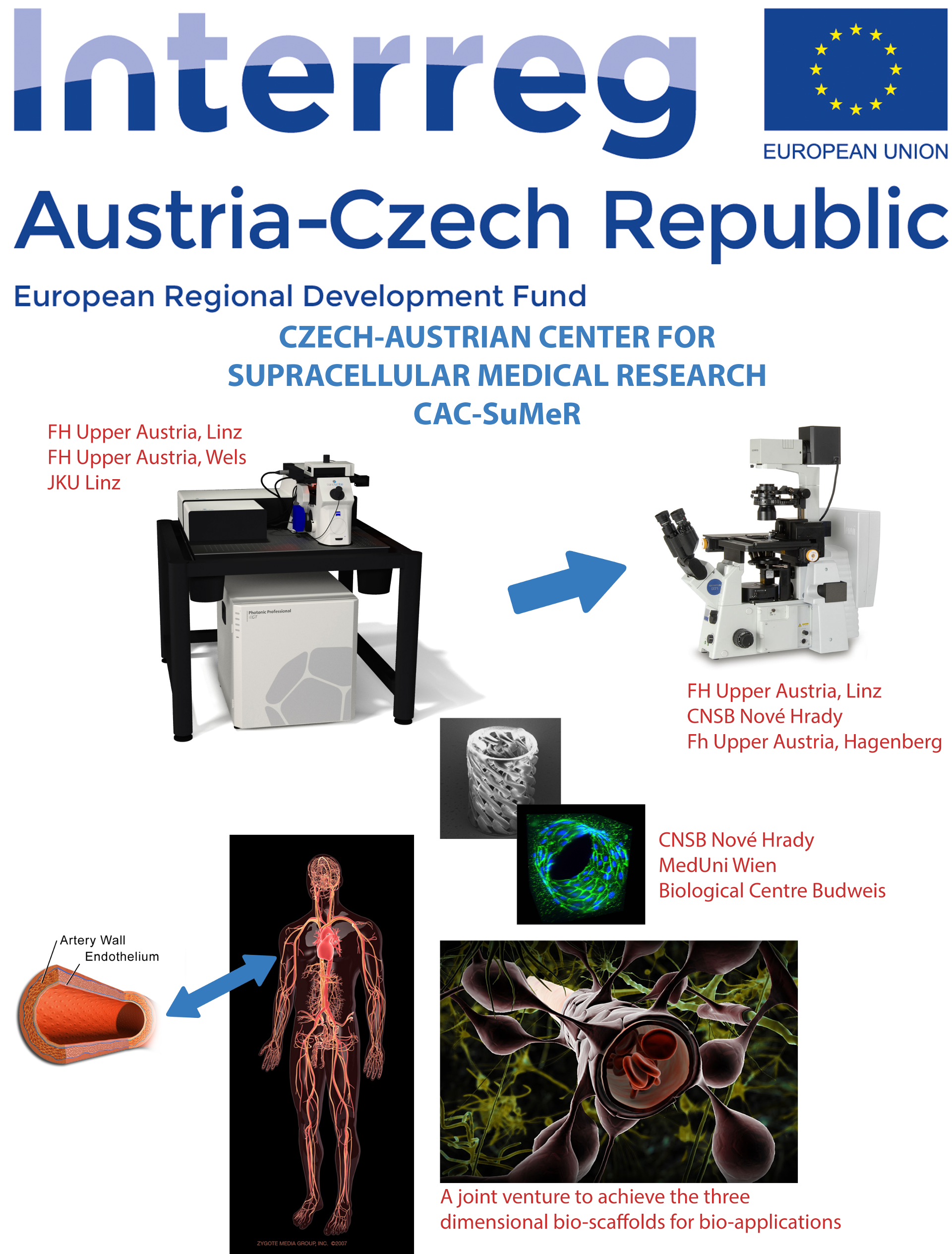
Zámek 136, 373 33 Nové Hrady
Research
Development of two-photon polarization microscopy into a tool of structural biology
Two-photon polarization microscopy (2PPM), a microscopy technique developed by our laboratoryallows sensitive observations of molecular processes taking place in living cells and animals. The technique works through observing changes of orientation of a fluorescent label (such as a fluorescent protein), attached to the protein of interest. 2PPM allows detecting changes in conformation of fluorescently labeled proteins, and also changes in interactions of fluorescently labeled proteins with other proteins. The technique has been successfully used for observing activation of G-proteins, changes in intracellular calcium orientation, changes in cell membrane voltage and other processes. Although the technique currently allows observing changes in orientation of the fluorescent label, it does not currently allow determining the orientation of the fluorescent label (fluorescent protein). We are now working on filling the gap, by studying optical properties of fluorescent molecules (fluorescent dyes such as F2N12S, fluorescent proteins) and finding ways to determine their orientation in synthetic lipid membranes and membranes of living cells by 2PPM.
Development of novel genetically encoded optical probes of molecular processes
2PPM takes advantage of anisotropic optical properties of fluorescent proteins in order to observe changes in conformation or protein-protein interactions of proteins of interest. The technique is applicable to many existing constructs consisting of a membrane protein labeled by a fluorescent protein, and often, 2PPM allows existing constructs to be used as optical probes of molecular processes. However, it should be possible to develop genetically encoded optical probes specifically for use in 2PPM. Such probes should have considerable advantages over probes used in techniques such as FRET, largely due to 2PPM's need for only a single fluorescent label. 2PPM probes should allow studies of molecular systems closer to natural state and simultaneous observations of multiple processes (multiplexing). Furthermore, because of 2PPMs ability to yield insights into protein structure, it should be possible to develop such probes in a rational, structure-guided fashion. We are currently pursuing development of various 2PPM probes of G-protein/GPCR activation and other processes.
Development of a voltage sensitive fluorescent protein / Organization of the mammalian odorant and pheromone detection systems
The brain is an electric organ. Therefore, being able to observe changes in membrane voltage of neurons is crucial for understanding of how the brain works. While it is possible to monitor electrical activity of neurons both directly (using electrodes) and indirectly (using voltage sensitive dyes), both approaches have severe limitations. Electrophysiological methods allow monitoring of only a few cells at a time. Voltage sensitive dyes have seen only limited applications due to their toxicity and low staining selectivity. Having a genetically encoded optical sensor of membrane voltage, a voltage sensitive fluorescent protein, would avoid the drawbacks of the current methods and allow detailed monitoring of complete neural circuits and neuronal assemblies. This in turn would allow us to decipher the organizing principles of the brain, as well as the particulars of individual brain subsystems. Our goal is to understand how the brain works. That is why we are working on developing a voltage sensitive fluorescent protein.
Recent publications:
Feng Y., Valley M.T., Lazar J., Yang A.L., Bronson R.T., Firestein S.J., Coetzee W.A., Manley J.L.:
SRp38 Regulates Alternative Splicing and Is Required for Ca2+ Handling in the Embryonic Heart.
Developmental Cell 16: 528-538, 2009.
Lazar J.: A method for obtaining structural and functional information on proteins, based on polarization fluorescence microscopy, and a device implementing said method.
Patent #302233 of the Industrial Property Office of the Czech Republic (2010).
Lazar J., Bondar A., Timr S., Firestein S.J.:
Two-photon polarization microscopy reveals protein structure and function.
Nature Methods 8: 684-690, 2011.
Timr S., Bondar A., Cwiklik L., Stefl M., Hof M., Vazdar M., Lazar J., Jungwirth P.:
Accurate Determination of the Orientational Distribution of a Fluorescent Molecule in a Phospholipid Membrane.
Journal of Physical Chemistry B 118: 855-863, 2013.
Bondar A., Lazar J.:
Dissociated G GTP and G Protein Subunits Are the Major Activated Form of Heterotrimeric Gi/o Proteins.
Journal of Biological Chemistry 289: 1271-1281, 2014.
Lazar J.:
A method for obtaining structural and functional information on proteins, based on polarization fluorescence microscopy.
Patent #8,643,177, United States Patent and Trademark Office (2014).
Han Z., Jin L., Chen F, Loturco J.J., Cohen L.B., Bondar A., Lazar J., Pieribone V.:
Mechanistic Studies of the Genetically Encoded Fluorescent Protein Voltage Probe ArcLight.
PLoS One 9: e113873, 2014.
Timr Š, Brabec J, Bondar A, Ryba T, Železný M, Lazar J, Jungwirth P.:
Nonlinear Optical Properties of Fluorescent Dyes Allow for Accurate Determination of Their Molecular Orientations in Phospholipid Membranes.
J Phys Chem B. 2015 Jul 30;119(30):9706-16
Links